A digital model of every patient, built from their own scans.
LivGemini combines artificial intelligence with physics to turn routine medical images into quantitative models of each patient's cardiovascular system. Our first product, VenusX, applies this to the thoracic aorta.
Images show the anatomy. Physics shows how it behaves.
- For clinicians
Quantitative 3D models from the scans you already acquire, and model-based estimates of what the images cannot show.
- For research teams
A way to study anatomy, blood flow and tissue biomechanics on real patient cohorts, with methods that are published and traceable.
- For device makers
Patient-specific anatomies and simulations to understand how devices meet real anatomies, before and after they reach patients.
AI for the anatomy. Physics for flow and mechanics.
Each model is built in three layers. We keep apart what is measured on the images, what is estimated by a model, and what the clinician decides.
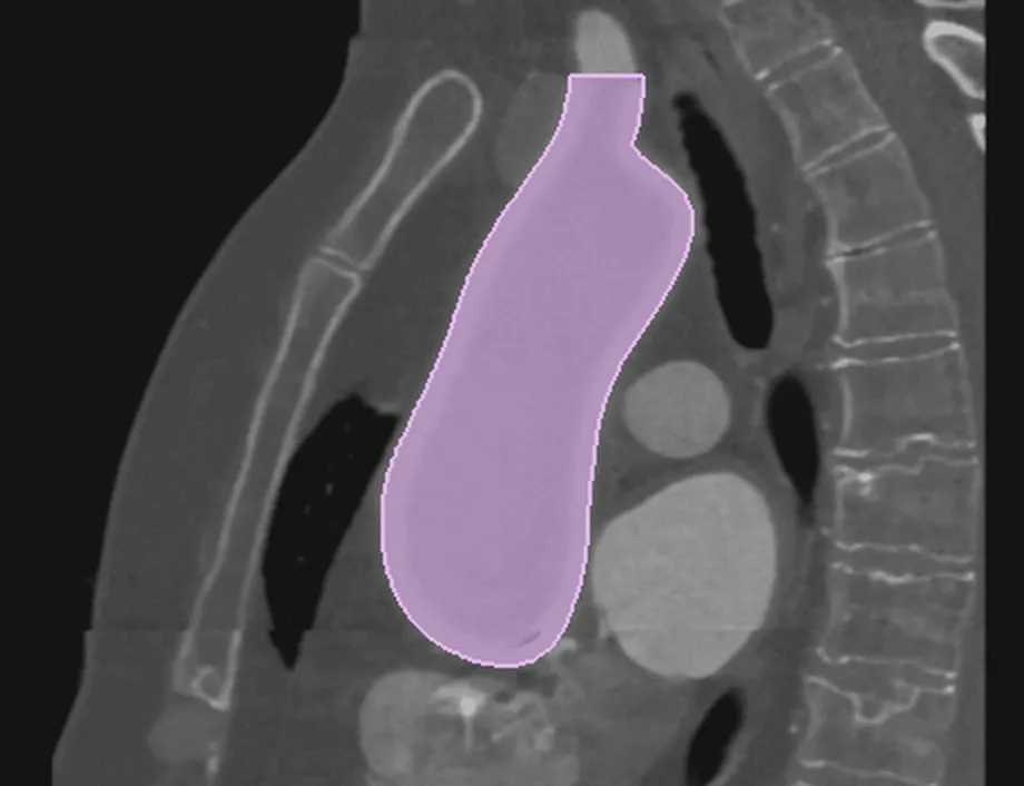 01AI · anatomy
Reconstruct the anatomy
Neural networks turn the patient's scan into a 3D model of the anatomy. A clinician reviews it before anything is computed on top.
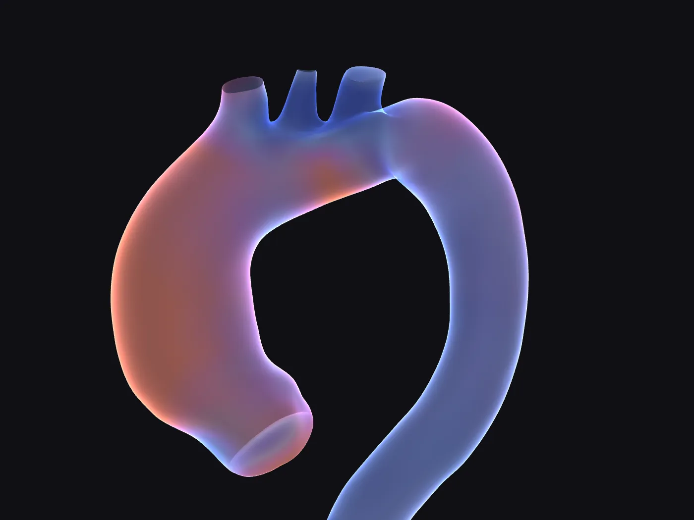 02Morphology
Assess its shape
Dimensions, curvature, volumes and motion describe each anatomy in numbers, and place it among other patients.
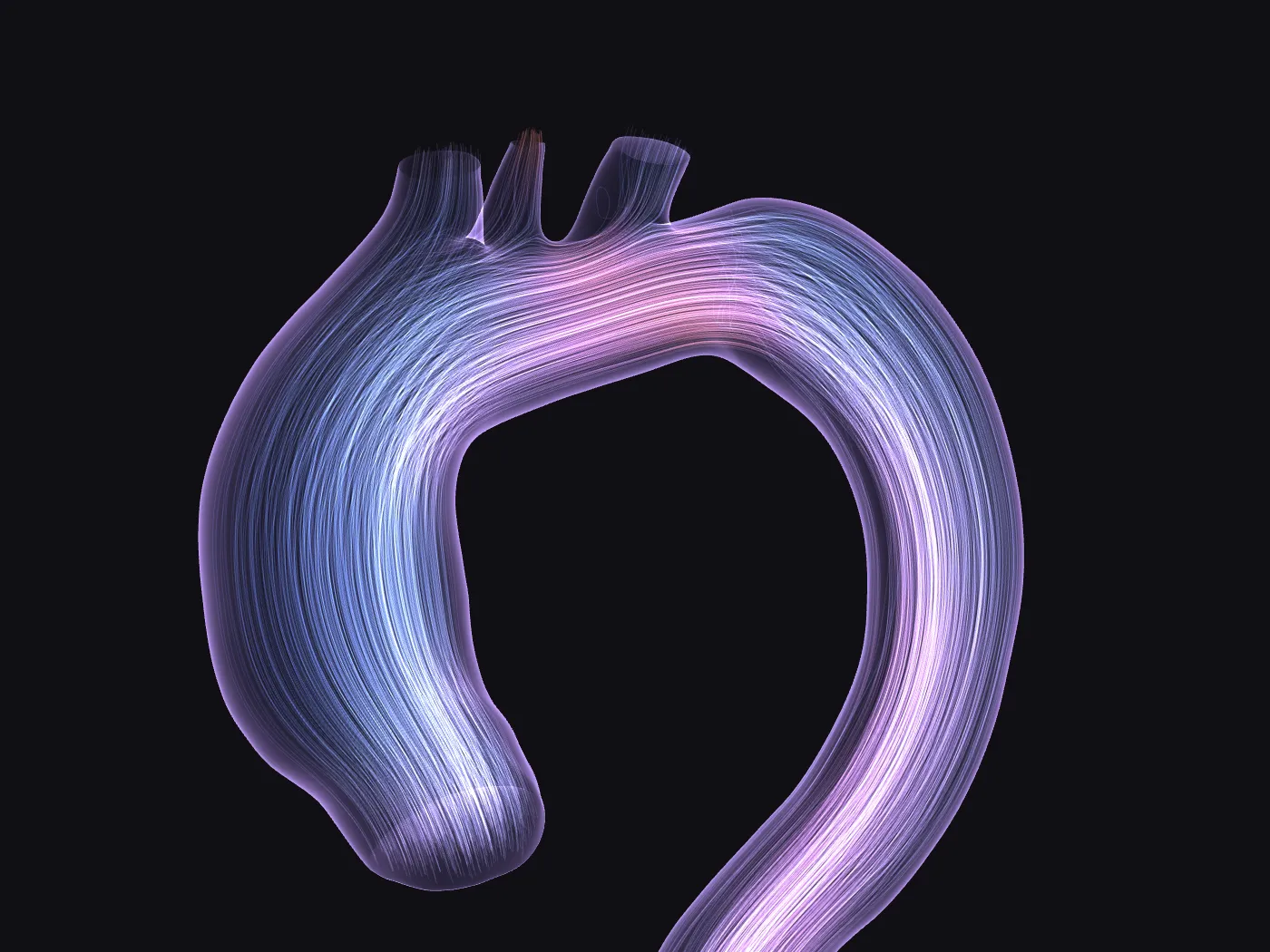 03Physics · simulation
Simulate what images cannot show
Physics-based models estimate blood flow, the biomechanics of tissues and walls, or the effect of a procedure. AI-built reduced-order models make them fast enough for a workstation.
The same method, across the cardiovascular system.
Thoracic aorta: segmentation, centerline and diameters from CT in VenusX Sizing. Flow estimates in VenusX Flow, in development.
Cardiac anatomy and motion, aneurysm growth from shape, endovascular planning, wall and valve mechanics, and virtual patient populations for device testing.

The thoracic aorta, from CT to report.
VenusX is a multiplatform desktop application for radiologists, surgeons and cardiologists who assess the thoracic aorta. It runs on Windows and macOS, processes images on the local computer and keeps the user in control of every automatic step.
Explore VenusX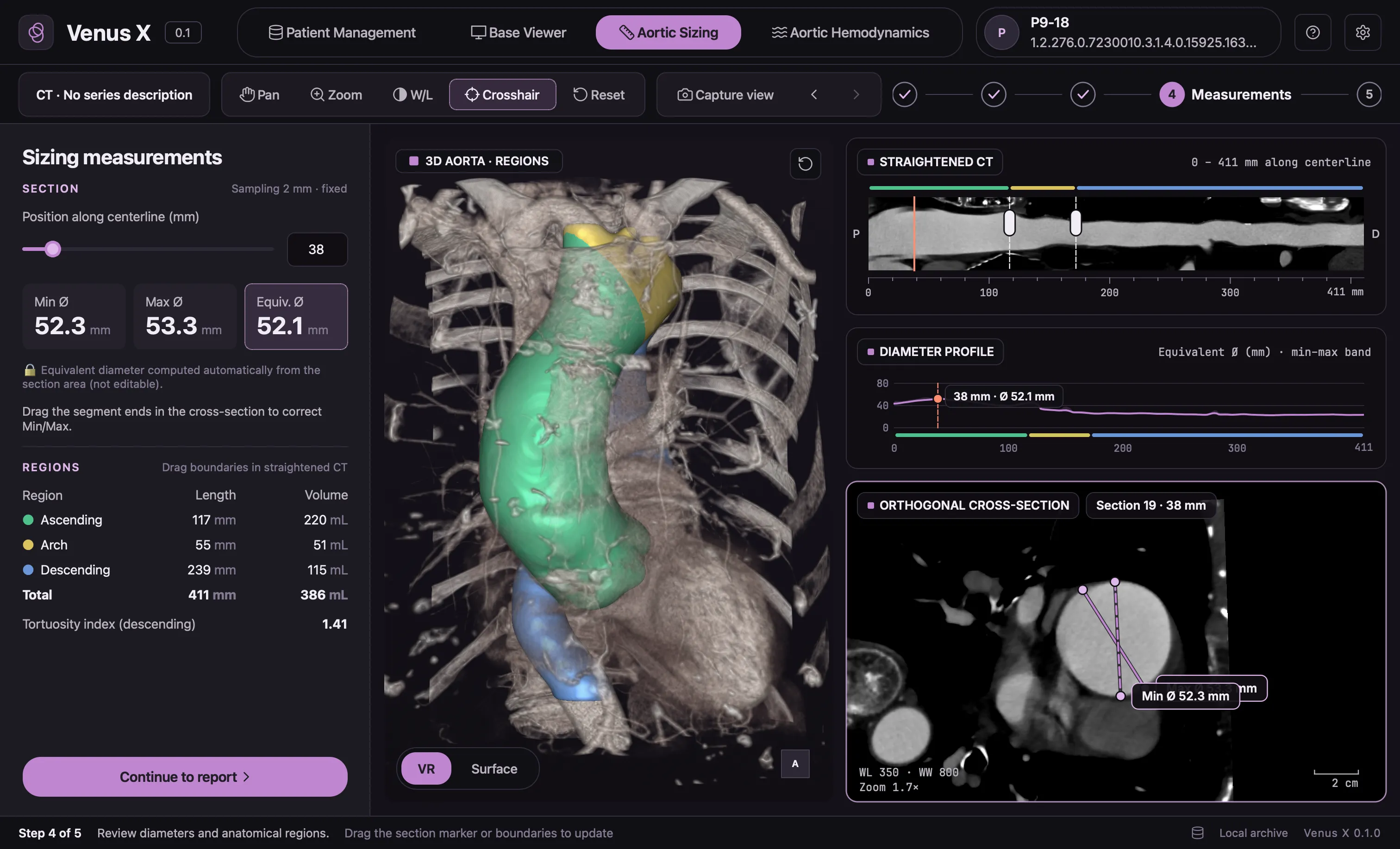
VenusX is not CE marked or FDA cleared. It is available for research use only and must not be used for clinical decision-making.
Published science, a certified process.
Our methods come from years of research in computational biomechanics. We state what is available, what is in development and what is still research.
- Peer-reviewed papers behind the methods
- 9
- Certified quality management system
- ISO 13485
- Hospitals in research collaborations
- 3
- CHU DijonClinical research collaboration
- Policlinico Tor VergataClinical research collaboration
- Massy HospitalClinical research collaboration
- ConfinisRegulatory partner
“It is a pleasure to collaborate with LivGemini and continue establishing new, innovative techniques for diagnosing and predicting aortic pathologies.”
 Prof. Jean-Philippe Verhoye MDVice-President, French Society of Cardiovascular and Thoracic Surgery · LivGemini clinical advisor
Work with us.
Evaluate VenusX
See VenusX Sizing on an anonymised case and discuss a research study on your own data.
Clinical collaboration Device manufacturersBuild a partnership
Bring VenusX to the teams using your devices, or use our modelling for your development.
Industrial partnership Investors and talentMeet the team
Who we are, the projects that fund our research, and the roles we are hiring for.
About LivGemini