01Input · imaging
Start from routine imaging
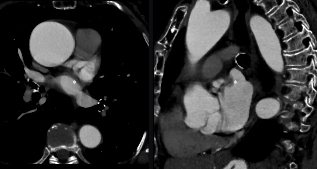
Models are built from the scans patients already have. CT is our starting point; time-resolved CT and other modalities add motion and function. No extra acquisition, no dedicated protocol.
We combine machine learning and physics. AI reconstructs the anatomy from imaging; physical laws describe how blood flows and how tissues and devices deform within it; AI again makes those physics fast enough to use outside a supercomputer.
Models are built from the scans patients already have. CT is our starting point; time-resolved CT and other modalities add motion and function. No extra acquisition, no dedicated protocol.
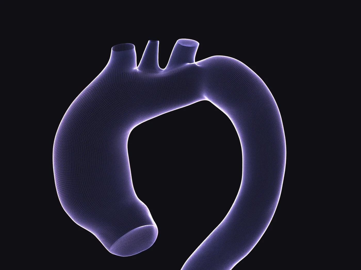
Neural networks turn the images into a 3D computational model of the anatomy: a surface and a volume mesh that keep the patient's own shape and are ready for measurement and simulation. The user reviews the result before anything is computed on it.
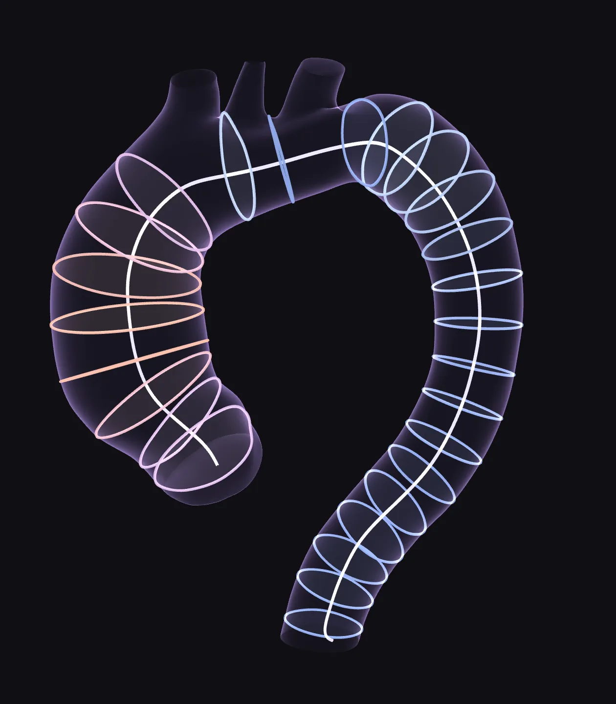
From the 3D model we describe the anatomy in quantitative terms: dimensions, curvature, volumes, how it moves over the cardiac cycle and how it compares with other patients. Which descriptors matter depends on the anatomy and on the clinical question.
Physics-based models compute what a scan cannot measure: blood flow, pressure and wall shear stress, the mechanics of the vessel wall, or the effect of a procedure or a device on the patient's anatomy. AI-built reduced-order models make these simulations fast enough for a workstation. Results are model estimates, and their reliability depends on the inputs.
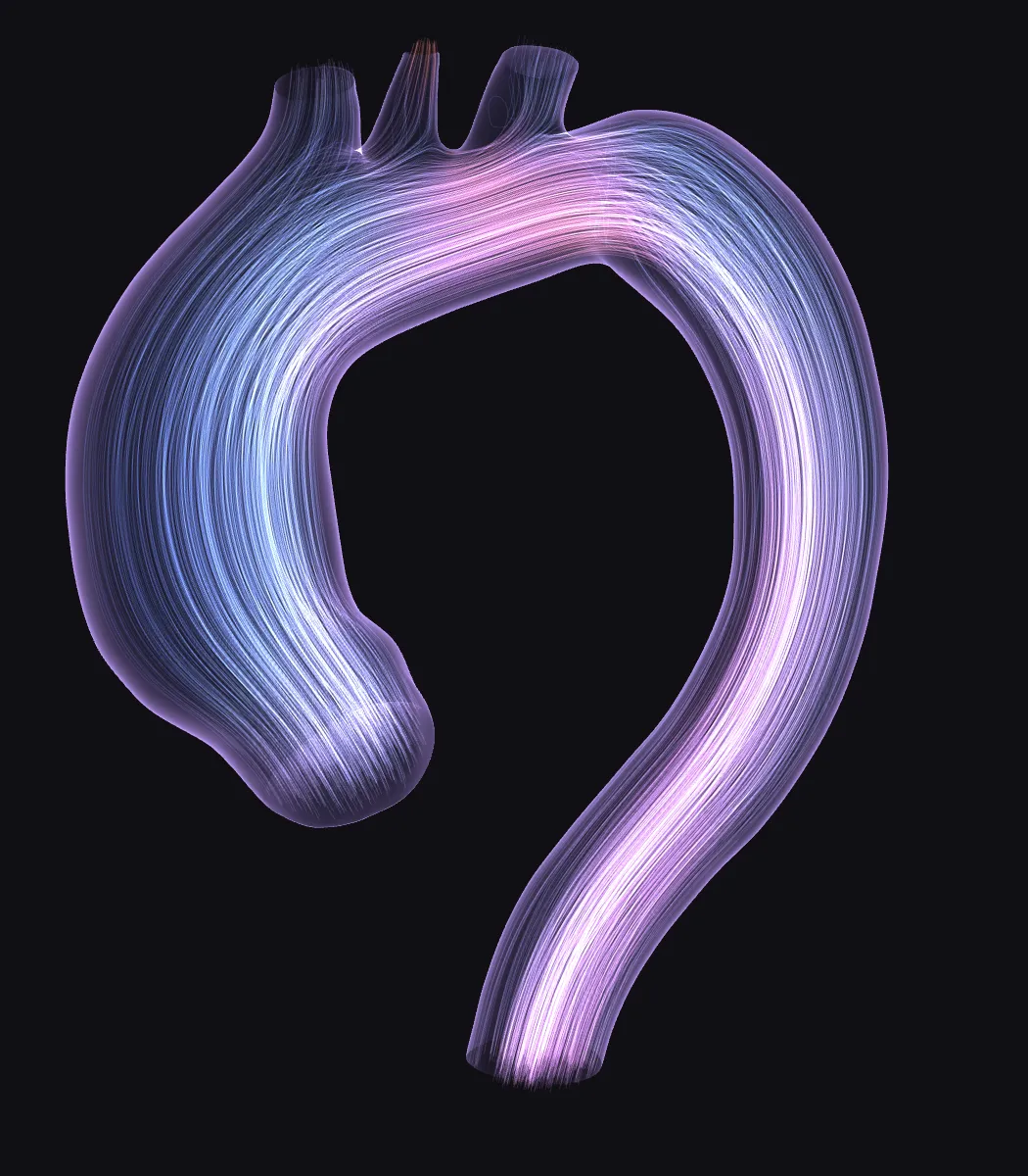
A model built on a patient's geometry is not automatically a full model of that patient's physiology. We say which quantities come from the images and which depend on assumptions.
High-fidelity flow and structural simulations take hours on dedicated hardware. That is fine for a study, not for a workstation in a hospital.
Neural networks reconstruct the anatomy. They run on the local computer, and the user reviews their output.
Established flow and structural solvers, with mesh morphing, generate reference simulations on many anatomies.
Those reference simulations train compact models that approximate the result for a new geometry in a fraction of the time.
VenusX is where the technology is already a usable product. The same methods are being developed for other questions; they are research, not products.
Sizing: segmentation, centerline, diameters and report, available for research evaluation. Flow: model-based hemodynamics, in development.
VenusXShape features beyond diameter, extracted from imaging, to study how fast an ascending aneurysm grows.
Reduced-order models to support endovascular planning and intra-operative navigation.
Fluid–structure models of vessel walls and heart valves, including heart motion and growth over time.
Sets of realistic 3D anatomies to test devices in silico before physical prototyping.
The team behind VenusX runs computational projects for medical device manufacturers, researchers and clinicians: from patient-specific anatomies to virtual deployment and flow.
Automatic extraction of anatomical structures from CT and MRI.
Sets of realistic 3D anatomies for in-silico trials.
Simulate and refine a device before physical prototyping.
Flow and structural models built on individual anatomies.
AI and physics-based models of risk and disease progression.
The core of our modelling, from reconstructing the anatomy to making physics fast, is developed in-house and protected.
For research groups and companies interested in patient-specific modelling, in VenusX or beyond it.