Skip to content
VenusX

Thoracic aorta analysis from CT.

LivGemini's first product. For radiologists, cardiac and vascular surgeons and cardiologists who assess the thoracic aorta, and for the research teams working with them.

Request a research demo

VenusX is not CE marked or FDA cleared. It is available for research use only and must not be used for clinical decision-making.

VenusX Sizing

A measured aorta, checked by you.

From CT import to report. The software proposes each result; the user reviews it before moving on.

01 · Import and review

Read the scan in every plane.

Linked 2D views, 3D rendering and measurement tools. Any view can be captured into the report.
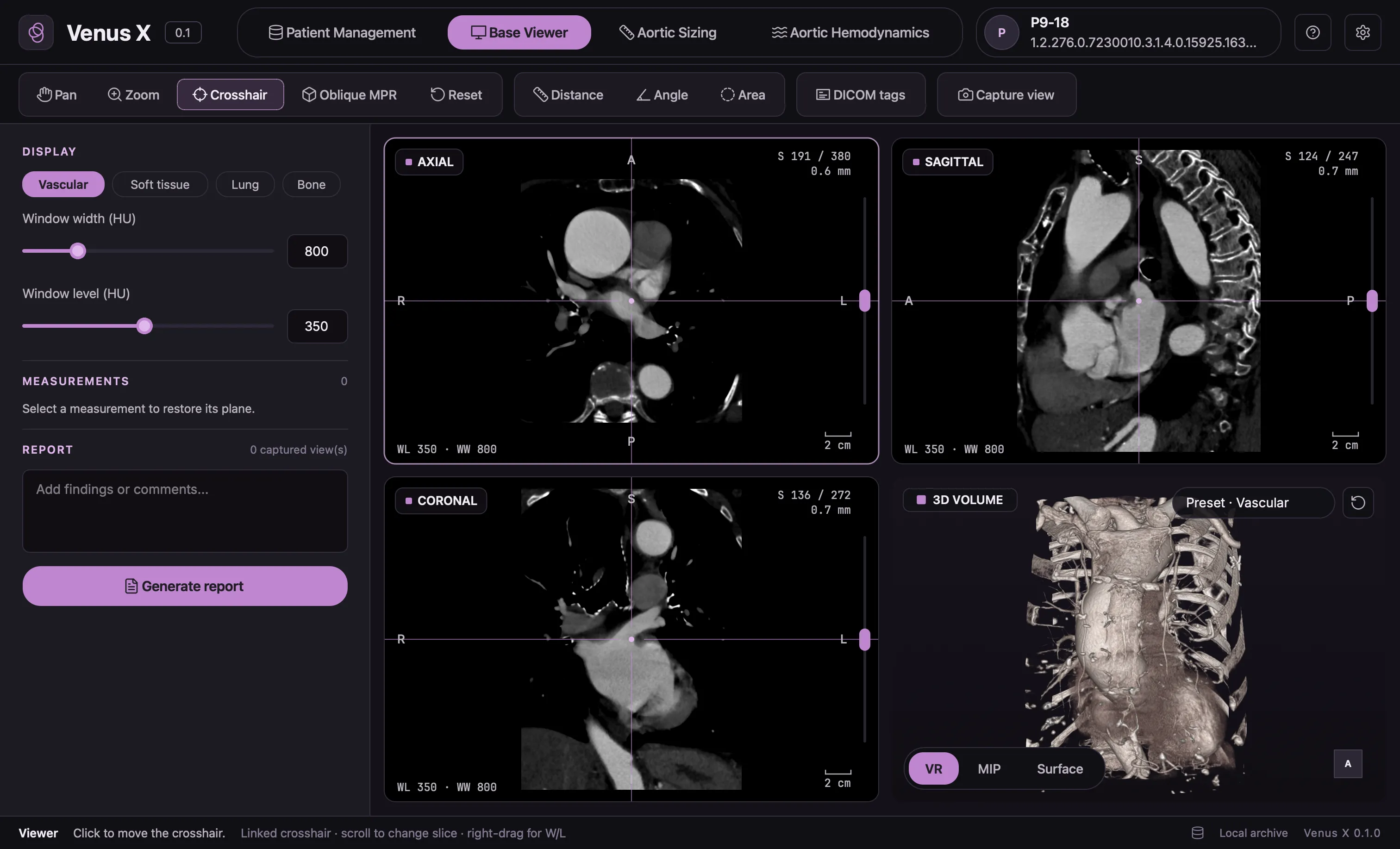
VenusX base viewer: axial, sagittal and coronal CT planes with a linked crosshair and a 3D volume renderingVenusX 0.1 · anonymised CT
The base viewer on an anonymised thoracic CT.
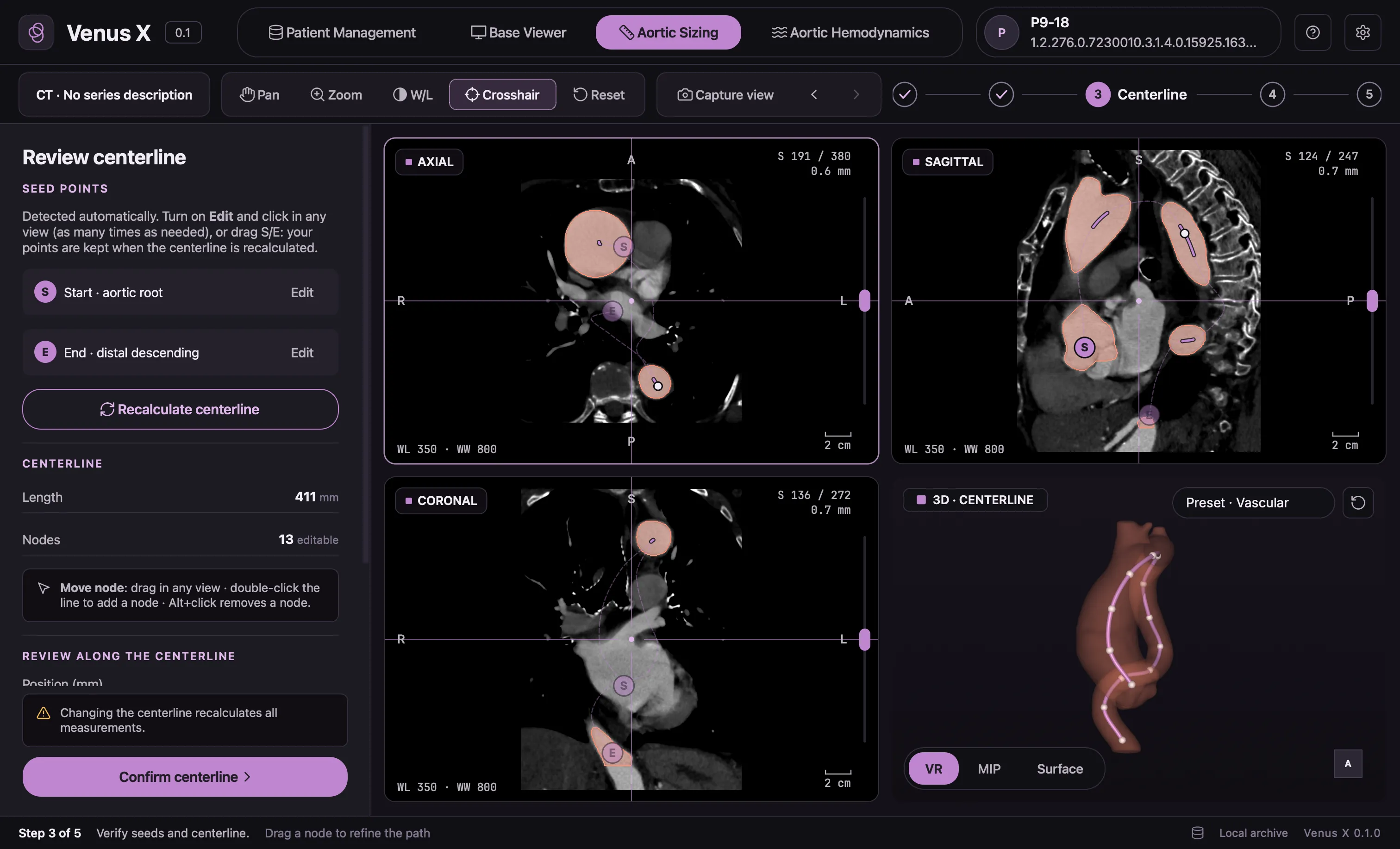
VenusX centerline review: seed points at the aortic root and distal descending aorta, lumen highlighted on CT planes, centerline nodes on the 3D aortaVenusX 0.1 · anonymised CT
Automatic results stay editable; measurements update with them.
02–03 · Segmentation and centerline

AI proposes, the user confirms.

An AI model segments the aorta and traces its centerline. The user reviews both before anything is measured.

04 · Measurements

From one section to the whole aorta.

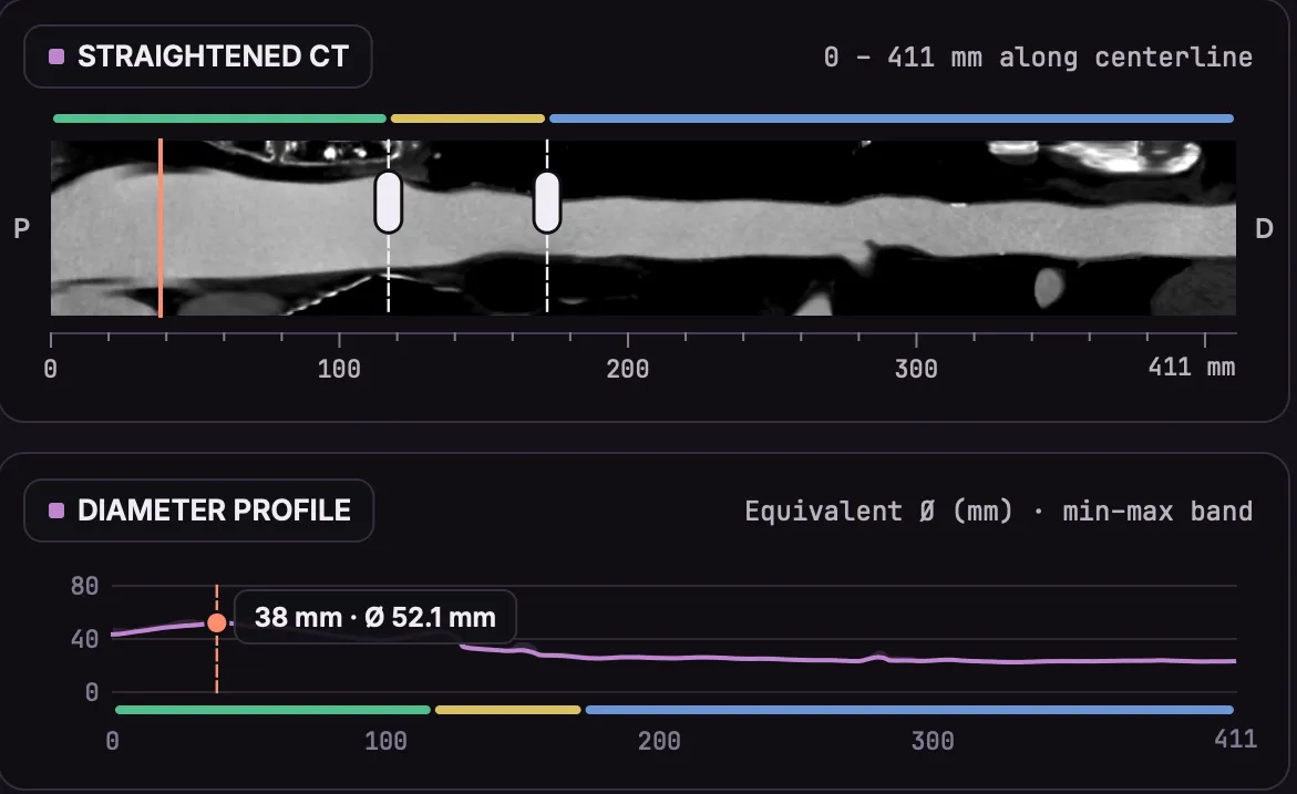
Diameters on sections perpendicular to the centerline, summarised by region with length, volume and tortuosity.
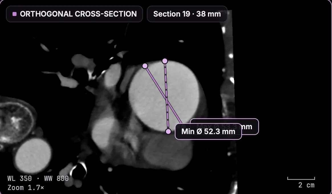
CT section perpendicular to the aortic centerline at 38 mm, with the minimum diameter of 52.3 mm drawn across the lumenSizing · orthogonal section
1 · Each section is cut perpendicular to the vessel and measured.
Straightened CT of the aorta over 411 mm with the diameter profile below it, the selected section marked at 38 mm with 52.1 mmSizing · diameter profile
2 · Together, the sections give the diameter profile of the whole aorta, region by region.
Sizing report
PDF
Summary
Max diameter
Length
Volume
Measurements by region
Ascending
Arch
Descending
Diameter profile
Key views
Comments and traceability
What a Sizing report contains: summary, measurements by region, diameter profile, key views, comments and the software version used.
05 · Report

A report the aortic team can read.

A structured PDF with measurements by region, captured views and comments, and the software version used.

VenusX Flow · in development

An estimate of the flow inside the same aorta.

Flow starts from the geometry confirmed in Sizing and the boundary conditions set by the user, and estimates wall shear stress, pressure and velocity.

The geometry comes from the patient's CT. Inflow, outflow split and vessel resistance are entered by the user or assumed, so the results are model estimates and depend on these inputs.
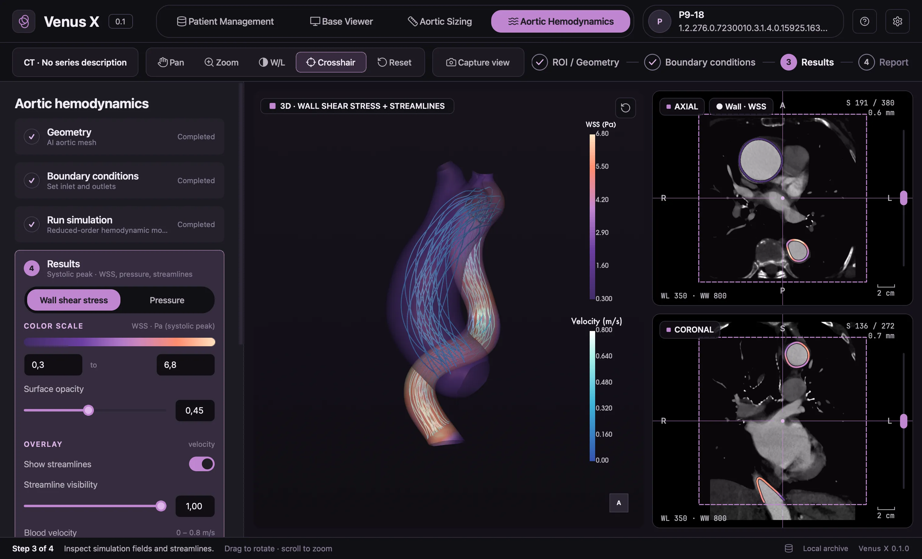
VenusX Flow development build: simulated wall shear stress in pascal and velocity streamlines in metres per second on the 3D aorta, with axial and coronal CTVenusX Flow · development build · simulation
Development build. Simulated values for illustration, not for clinical use.
At a glance

What goes in, what comes out.

Input
Thoracic CT series in DICOM, imported from PACS, a local folder, a ZIP file or removable media.
User checks
Every automatic step: segmentation, centerline, regions and measurements.
Sizing output
Diameters along the centerline; length, volume and tortuosity by region; captured views; PDF report.
Flow output
Simulated wall shear stress, pressure and velocity on the 3D aorta. In development.
Platforms
Multiplatform desktop application for Windows and macOS workstations and laptops. All processing runs locally.
Access
Research evaluation with clinical and academic centres, under an agreement with LivGemini.
Under the hood

VenusX runs on LivGemini's modelling technology.

AI reconstructs the aorta, geometry gives the measurements, and reduced-order physics models estimate the flow. The technology page explains each layer, what is measured and what is assumed, and how we build and test the software.

How the technology works
FAQ

Questions we hear.

Can VenusX be used for patient care?

VenusX is not CE marked or FDA cleared. It is available for research use only and must not be used for clinical decision-making.

Do images leave the hospital?

No. VenusX processes the CT on the computer where it is installed. Our verification includes a check that no network connection is opened during a complete analysis.

How much of the workflow is automatic?

Segmentation, centerline detection and measurements are computed automatically. The user reviews and confirms each step, can edit the centerline and correct diameters, and decides what goes into the report.

Which CT protocols are supported?

VenusX needs an axial CT series with at least three slices. The protocols validated for each module are listed in the documentation we share for a research evaluation.

Is VenusX Flow available?

VenusX Flow is in development and evaluation. We show it in demos as a separate module and discuss its use in research projects case by case.

Where are the instructions for use?

The electronic instructions for use are available on request from support@livgemini.com.

See VenusX Sizing on an anonymised case.

A live walkthrough from CT import to the PDF report, for clinical and research teams evaluating the software for research use.
Important notice

Who is visiting?

Some content on this website concerns medical devices and software that require specialised expertise. It is intended for healthcare professionals and for professionals working in the medical technology field (for example researchers, engineers, regulatory and industry experts).

Please select the option that describes you:

If you are a patient or member of the public, please consult your physician for medical advice. Nothing on this website constitutes medical advice.