Skip to content
← All publications
Image segmentationSupports VenusX Sizing

Segmentation of the aorta in systolic phase from 4D flow MRI: multi-atlas vs. deep learning

Magnetic Resonance Materials in Physics, Biology and Medicine
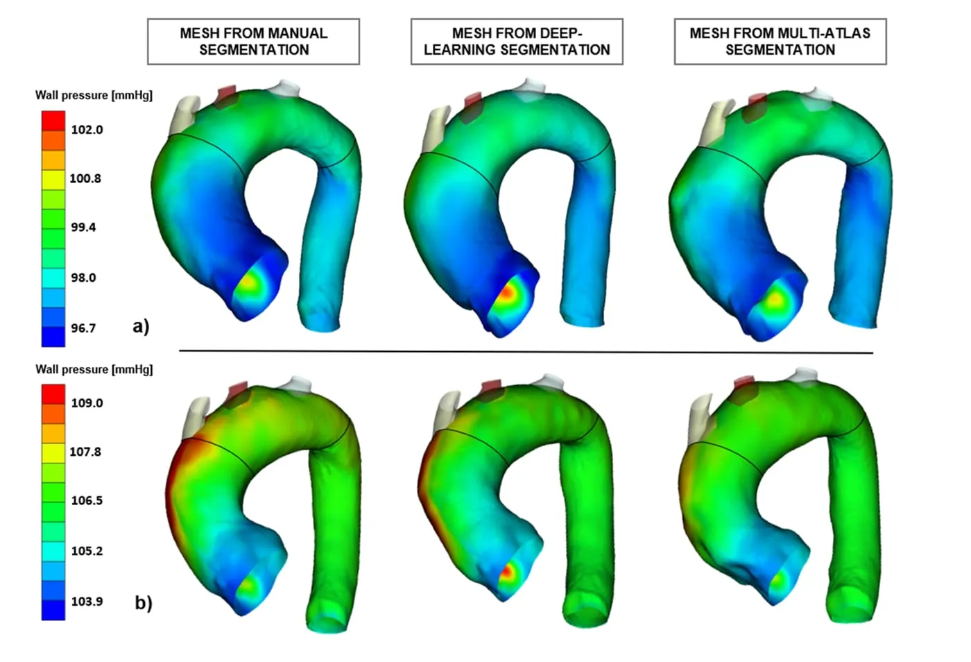
Figure from “Segmentation of the aorta in systolic phase from 4D flow MRI: multi-atlas vs. deep learning”

Question

How do atlas-based and deep learning methods compare for automatic aortic segmentation?

What the paper shows

Deep learning and multi-atlas segmentation were compared on 36 patients, both on the segmentation itself and on the wall pressure computed from each model. Deep learning gave the more accurate segmentation and the pressures closest to those from manual segmentation.

Authors

Marin-Castrillon D. M., Geronzi L., Boucher A., Lin S., Morgant M.-C., Cochet A., Rochette M., Leclerc S., Ambarki K., Jin N., Aho L. S., Lalande A., Bouchot O., Presles B.
Read the full paper

Study VenusX with us.

We co-design studies with clinical and academic centres. Data are shared only under an agreement with your institution.

Important notice

Who is visiting?

Some content on this website concerns medical devices and software that require specialised expertise. It is intended for healthcare professionals and for professionals working in the medical technology field (for example researchers, engineers, regulatory and industry experts).

Please select the option that describes you:

If you are a patient or member of the public, please consult your physician for medical advice. Nothing on this website constitutes medical advice.